abnormal thyroid cancer ultrasound colors
Intranodular vascularity and high RI indices were the specific Doppler signs for malignant thyroid nodules. Buy 2021 Quality Abnormal Thyroid Cancer Ultrasound Color Doppler directly with low price and high quality.

Intranodular Vascularity Grading On Color Doppler And Representative Download Scientific Diagram
The thyroid is imaged using a high-frequency linear transducer 715 MHz with the patient lying supine with the neck in hyperextension.

. Flow should be readily seen scattered throughout but not dominate the gland or have abundant aliasing. Better penetration lower resolution. These images are examples of pathology I detect with sonograms.
It is generally normal unless there is too much color which would have been mentioned in the report. Im not an ultrasound tech so Ill answer to the best of my knowledge. Since Gray scale and Doppler have their own strengths and weaknesses they were complementary rather than competitive modalities in diagnosing benign from malignant thyroid nodules.
What is more up to 24 of papillary thyroid carcinomas may have a halo be it complete or incomplete 2 Color Doppler intranodular flow usually malignant. While a halo around a well-marginated hypoechoic or isoechoic nodule is typical of a follicular adenoma 3 it is absent in 50 of benign nodules 2. Ultrasound probe both emits and receives sound waves.
Corinne Deurdulian answered Radiology 23 years experience Blood flow. Normal ultrasound of thyroid vascularity using colour doppler. However less than 7 of thyroid nodules are malignant.
There was a significant association between Resistive Index and Pulsatility Index with malignancy with a cutoff of RI 0715 P0005 and PI 0945 P0007 respectively. Color on your thyroid ultrasound means that color doppler was applied and blood flow was detected. I don t think it has a color cancer cannot be actually diagnosed by ultrasound but it can show a mass that is either fluid filled or solid.
High-resolution ultrasonography US is commonly used to evaluate the thyroid gland but US is frequently misperceived as unhelpful for identifying features that distinguish benign from malignant nodules. Thyroid nodular disease is one of the most frequent endocrine pathologies in everyday practice. The combination of calcification RI 0715 with PI 0945 had a very diagnostic yield for diagnose of malignancy PPV 666 and NPV 984.
Lower penetration higher resolution. Use a curvi-linear ultrasound probe to accurately measure or visualise a retrosternal thyroid. In contrast other studies have shown that ultrasound features such as coarse calcifications more tall than wide irregular borders and increased blood flow within the nodule can be helpful to identify thyroid cancer.
In contrast other studies have shown that ultrasound features such as coarse calcifications more tall than wide irregular borders and increased blood flow within the nodule can be helpful to identify thyroid cancer. Ultrasound Sonographic appearance depends on underlying etiology and may include. Other links to color or gray scale images will be included here later.
Aneurysms arteriosclerosis obstruction dissection. 2D black and white image in 1mm slice. 48k views Answered 2 years ago Thank 10 thanks Similar questions.
Since the late 1960s ultrasound has become essential in the examination of the thyroid gland with the increased availability of high-frequency linear array transducers and computer-enhanced imaging capabilities of modern day portable ultrasound equipment in a clinic- or office-based. If its fluid filled its generally classified as a cyst. The first links in each row here correspond to ultrasound color post-processed images.
Furthermore the components of ultrasound of the thyroid gland are well established including the appearance of the normal thyroid gland and thyroid pathology. Abnormal size of the thyroid gland alteration in thyroid echotexture may be diffuse or nodular abnormal color flow Doppler patterns Treatment and prognosis. Thyroid nodules are common and occur in up to 50 of the adult population.
Definition general. 2 - 20 MHz. Answer 1 of 3.
The use of ultrasound for thyroid cancer has evolved dramatically over the last few decades.

Color Doppler Patterns A Pattern 0 Normal Thyroid Vascularity B Download Scientific Diagram

Benign Thyroid Nodule With Large Intratumoral Cyst Transverse Download Scientific Diagram

3 D Ultrasound And Thyroid Cancer Diagnosis A Prospective Study Ultrasound In Medicine And Biology

Role Of Color Doppler Us A Transverse Gray Scale Image Of Download Scientific Diagram

Abnormal Jugular Vein Detected During Thyroid Ultrasound

Normal Thyroid Color Doppler Youtube

Thyroid Scintiscanning An Overview Sciencedirect Topics
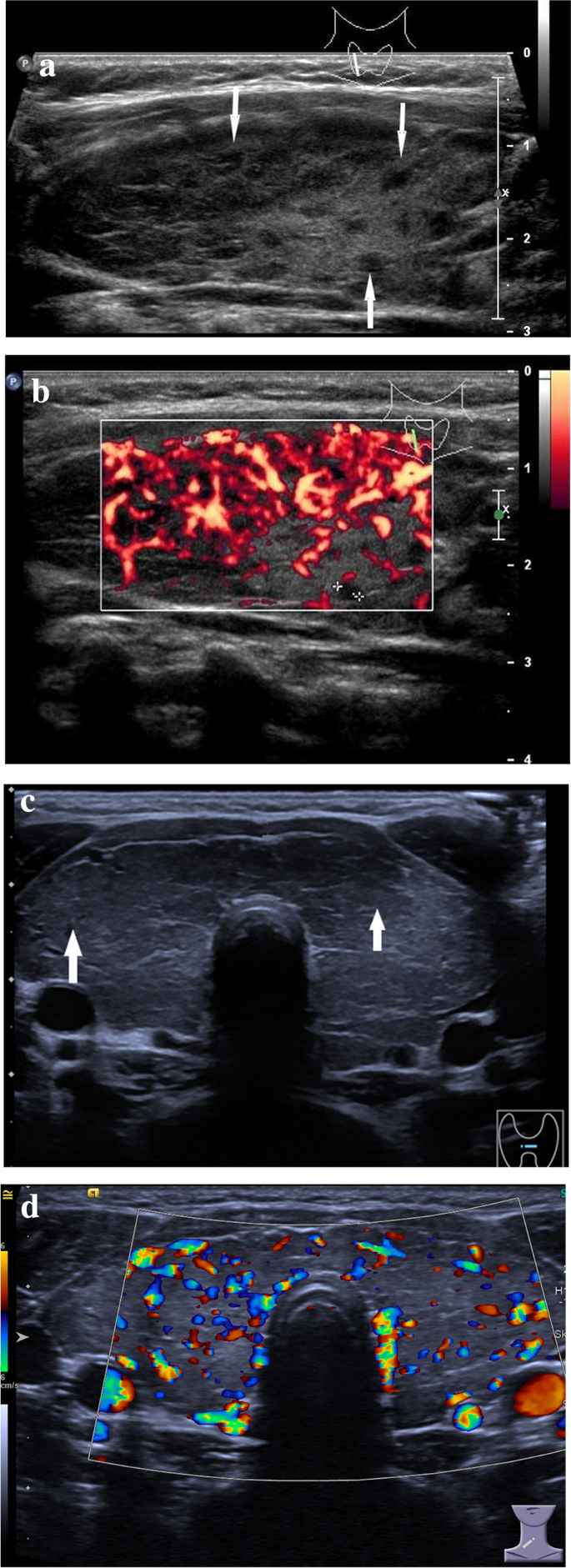
Ultrasound Findings Of The Thyroid Gland In Children And Adolescents Springerlink

Multinodular Goiter Transverse Gray Scale Ultrasound A And Color Download Scientific Diagram
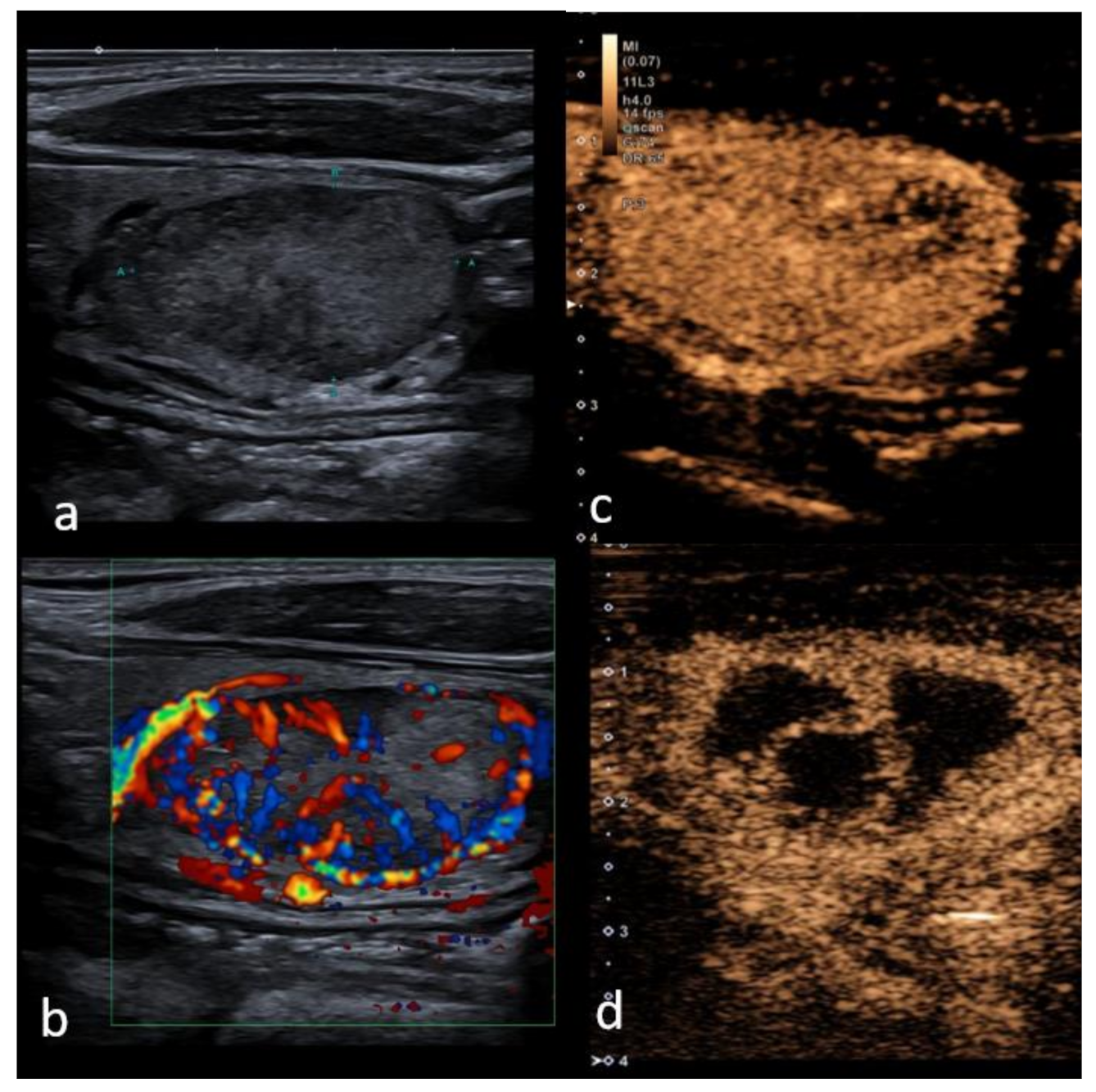
Cancers Free Full Text Performance Of Contrast Enhanced Ultrasound In Thyroid Nodules Review Of Current State And Future Perspectives Html

Ultrasound Of The Neck There S More To See Than Thyroid Nodules Youtube

Intranodular Vascularity Grading On Color Doppler And Representative Download Scientific Diagram

Hashimoto S Thyroiditis Transverse Gray Scale Ultrasound A And Color Download Scientific Diagram

Role Of Color Doppler Us A Transverse Gray Scale Image Of Download Scientific Diagram

Ultrasound Findings Of The Thyroid Gland In Children And Adolescents Springerlink
Benign And Suspicious Ultrasound Features And Pathological Findings He Download Scientific Diagram
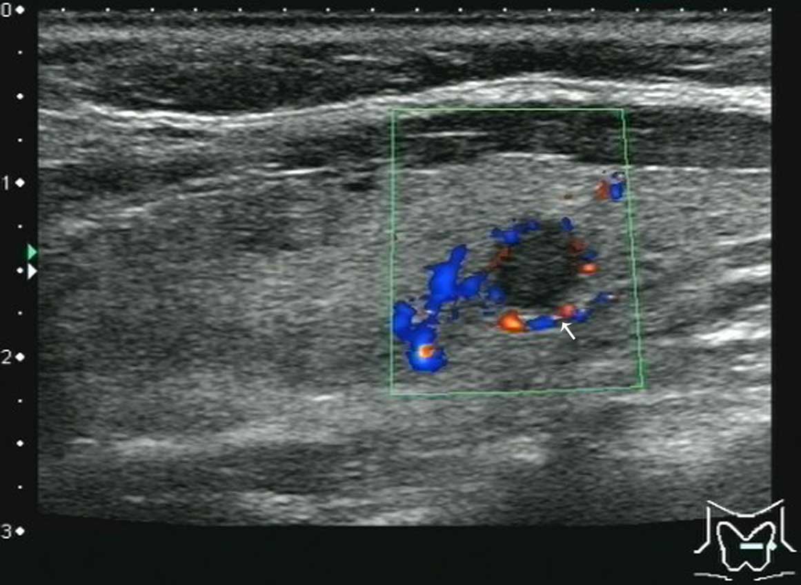
Ultrasonic Features Of Papillary Thyroid Microcarcinoma Coexisting With A Thyroid Abnormality
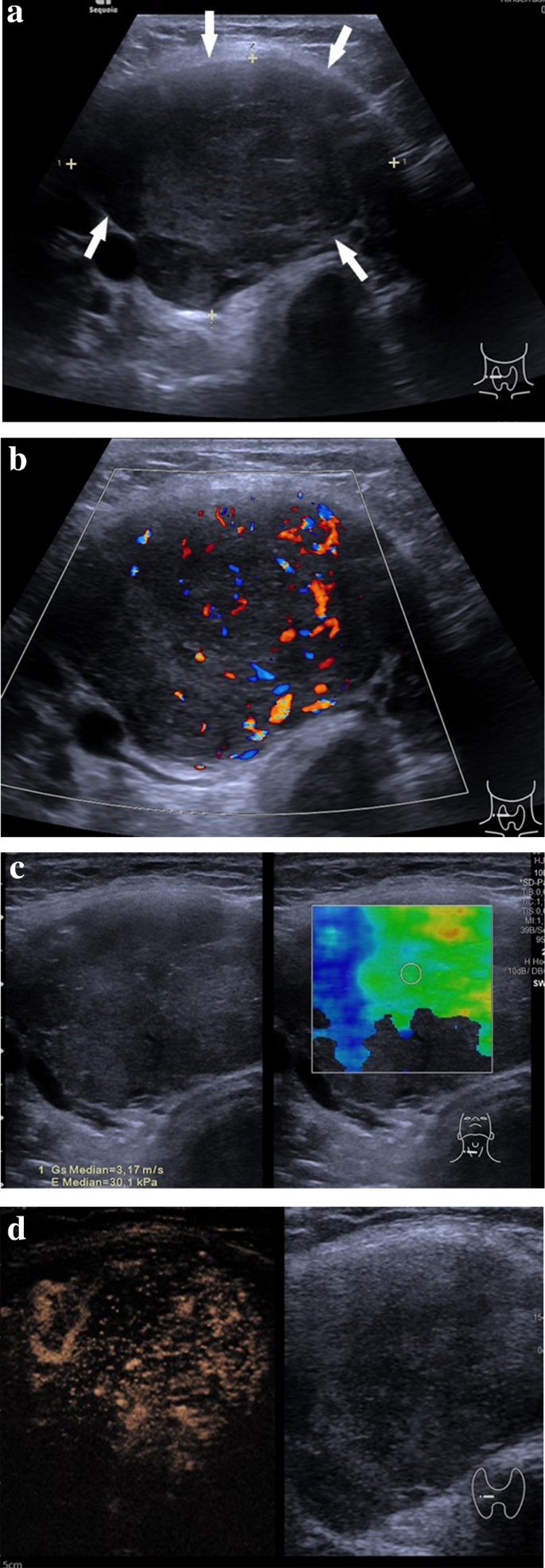
Ultrasound Findings Of The Thyroid Gland In Children And Adolescents Springerlink
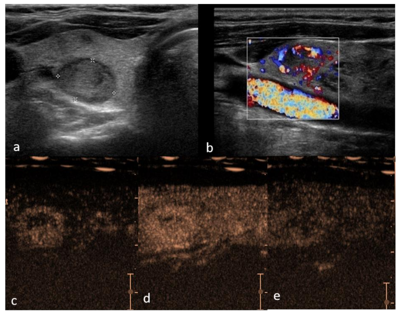
Cancers Free Full Text Performance Of Contrast Enhanced Ultrasound In Thyroid Nodules Review Of Current State And Future Perspectives Html